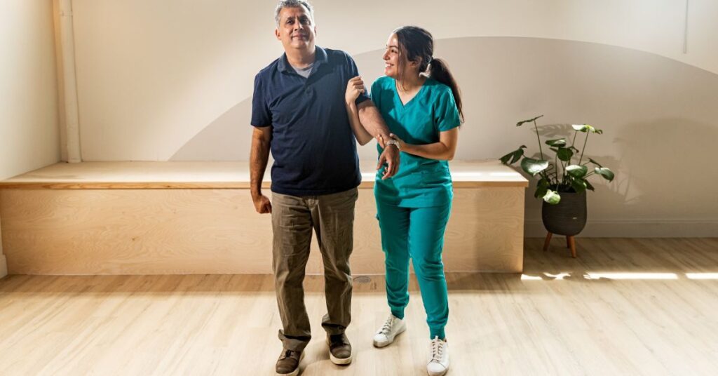
A cerebrovascular accident (CVA), commonly known as a stroke, alters the trajectory of life in an instant. Whether caused by an ischemic blockage interrupting blood flow to brain tissue or a hemorrhagic rupture causing localized bleeding, a stroke disrupts the delicate neural circuitry that governs motor control, sensory processing, balance, speech, and spatial orientation. For stroke survivors and their families, the immediate post-acute period is often filled with profound uncertainty, emotional distress, and overwhelming questions about the future.
Among all the inquiries presented to multidisciplinary medical teams, one fundamental question dominates every initial clinical consultation: “What actually happens during physiotherapy after a stroke in the first 3 months?”
The decision to focus on the first 90 days is not arbitrary; it is rooted in biological necessity. The first 12 weeks following a cerebrovascular event represent a unique, time-sensitive physiological window known as the hyper-acute and sub-acute recovery window. During this critical interval, the human brain exhibits its highest biological capacity for neuroplasticity—the innate cellular mechanism through which the central nervous system reorganizes its structural and functional connections to bypass damaged tissue.
At Rayara Kirana Physiotherapy & Rehab Center, our clinical philosophy is built around maximizing this 90-day window. Led by senior neuro-physiotherapists across Banashankari, Basavanagudi, and broader Bangalore, our team delivers structured, evidence-based neurological rehabilitation protocols designed to transition patients from acute bed-bound immobility to upright, functional independence.
This comprehensive, doctor-reviewed guide provides an exhaustive, month-by-month analysis of the rehabilitation journey, detailing the physiological changes, clinical milestones, therapeutic interventions, home safety strategies, and long-term care plans required during the first 3 months of stroke recovery.
The Biological Foundation: Why the First 90 Days Dictate Long-Term Recovery
To understand the structure of physiotherapy after a stroke in the first 3 months, one must first examine the cellular and neurological events that take place within the brain immediately following an ischemic or hemorrhagic insult.
Understanding Ischemic Injury and the Penumbra
When a cerebral artery is occluded, the core tissue at the center of the blockage suffers rapid cell death (infarction) within minutes. Surrounding this ischemic core lies a zone of compromised but potentially salvageable neural tissue called the ischemic penumbra. Emergency medical interventions—such as intravenous thrombolysis (tPA) or mechanical thrombectomy—aim to save the penumbra. Once the patient is medically stabilized, neurological rehabilitation takes over to stimulate the surviving neural tissue surrounding the lesion.
The Mechanism of Neuroplasticity
For decades, classical neurology operated under the assumption that the adult central nervous system was static and incapable of repair. Modern neurobiology has completely dismantled this myth. Neuroplasticity refers to the brain’s ability to alter its structure, function, and synaptic connections in response to internal stimuli, environmental demands, and repetitive motor learning.
During the first 3 months post-stroke, the brain undergoes a cascade of neuroplastic events:
Spontaneous Biological Recovery: In the initial days, systemic inflammation subsides, local brain edema clears, and uninjured neural pathways regain baseline metabolic function.
Synaptogenesis and Axonal Sprouting: Endogenous growth factors, such as Brain-Derived Neurotrophic Factor (BDNF), reach peak concentration in the peri-infarct zone. Neurons begin sprouting new dendritic spines and establishing new synaptic connections (synaptogenesis).
Unmasking Latent Pathways: The brain activates silent, pre-existing neural pathways that were previously inactive, rerouting motor signals from the motor cortex through alternative spinal tracts (such as the uncrossed anterior corticospinal tract).
Cortical Remapping: Undamaged areas of the cerebral cortex—including the premotor cortex and supplementary motor area—take over motor execution duties previously performed by the damaged region.
+-----------------------------------------------------------------------------------+
|                         THE NEUROPLASTICITY RECOVERY WINDOW                       |
+-----------------------------------------------------------------------------------+
| TIME WINDOW           | BIOLOGICAL STATE              | CLINICAL IMPLICATION     |
+-----------------------+-------------------------------+---------------------------+
| Months 1 to 3         | Peak BDNF expression, rapid   | Maximum capacity for      |
| (Sub-Acute Window)    | synaptogenesis, unmasking     | motor re-learning; high   |
|                       | of latent neural pathways.    | therapy intensity needed. |
+-----------------------+-------------------------------+---------------------------+
| Months 4 and Beyond   | Consolidation of synaptic     | Recovery continues via    |
| (Chronic Phase)       | maps; deceleration of         | high-repetition task     |
|                       | spontaneous neural growth.    | training & adaptation.    |
+-----------------------------------------------------------------------------------+
The Danger of Learned Non-Use and Maladaptive Plasticity
Neuroplasticity is a double-edged sword: it responds to both correct movement patterns and compensation strategies. If a stroke patient is left without structured physical therapy during the first 3 months, two dangerous phenomena occur:
Learned Non-Use: The patient relies entirely on their unaffected limbs to perform all daily tasks. Consequently, the cortical representation of the affected limb within the brain’s motor cortex shrinks, leading to permanent functional loss.
Maladaptive Plasticity: The patient adopts abnormal compensatory movements—such as severe hip hiking, lateral trunk leaning, or excessive shoulder elevation—to move. These abnormal movement patterns become hardwired into the brain’s motor map, making normal gait and arm function nearly impossible to restore later.
Structured, daily neurological physiotherapy during the sub-acute phase prevents maladaptive habits and drives recovery along anatomically correct pathways.
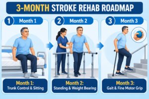
Month 1 (Weeks 1–4): Acute Bedside Rehab, Tone Management & Core Activation
The initial 30 days of physiotherapy after a stroke in the first 3 months represent the acute and early sub-acute phase. During this period, the patient transitions from the intensive care unit (ICU) or acute hospital ward to early bedside rehabilitation. The overarching clinical goal of Month 1 is not immediate unassisted walking, but preventing secondary immobility complications, managing altered muscle tone, protecting fragile joints, and re-establishing foundational core control.
Clinical Objective 1: Comprehensive Bed Positioning and Skin Integrity
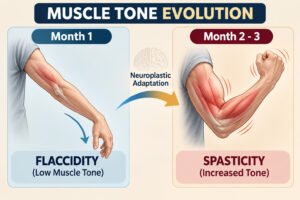
In the immediate aftermath of a stroke, the affected side typically enters a state of flaccidity (hypotonia)—a complete loss of voluntary muscle control and muscle tone. The limb feels heavy, limp, and powerless. Improper positioning during this flaccid state causes severe structural complications.
Preventing Decubitus Ulcers (Bedsores): Immobilized patients are at extreme risk of skin breakdown over bony prominences such as the sacrum, heels, greater trochanters, and scapulae. Physiotherapists establish strict 2-hour turning schedules, utilizing specialized pressure-relieving air mattresses and foam wedges.
Anti-Spastic and Anti-Edema Positioning: The stroke-affected arm is elevated on pillows above heart level to prevent fluid accumulation (dependent edema) in the hand. The hip and knee are supported in slight flexion to prevent external rotation contractures of the leg.
Passive Range of Motion (PROM) Exercises: Therapists perform daily, full-spectrum passive joint movements for the shoulder, elbow, wrist, fingers, hip, knee, and ankle. PROM keeps joint capsules lubricated, maintains muscle length, and provides crucial proprioceptive feedback to the recovering brain, mirroring safety principles detailed in our resource on structured physiotherapy for faster recovery after surgery.
+-----------------------------------------------------------------------------------+
|                        MONTH 1 CLINICAL FOCUS (WEEKS 1 TO 4)                       |
+-----------------------------------------------------------------------------------+
| PRIMARY DOMAIN        | CLINICAL TARGET               | THERAPEUTIC TECHNIQUE     |
+-----------------------+-------------------------------+---------------------------+
| Joint Protection      | Prevent shoulder subluxation  | Pillow support, therapeutic|
|                       | and ankle joint contractures. | slings, passive ROM.      |
+-----------------------+-------------------------------+---------------------------+
| Bed Mobility          | Independent rolling and       | Segmental trunk rolling,  |
|                       | bridging in bed.              | gluteal bridging drills.  |
+-----------------------+-------------------------------+---------------------------+
| Postural Control      | Symmetrical upright sitting   | Axial weight-shifting on  |
|                       | at the edge of the bed.       | bed edge, mirror feedback.|
+-----------------------------------------------------------------------------------+
Clinical Objective 2: Preventing Hemiplegic Shoulder Subluxation
One of the most painful and debilitating complications of early stroke recovery is shoulder subluxation—the partial dislocation of the humerus bone out of the glenoid cavity. Because the rotator cuff muscles (supraspinatus, infraspinatus, subscapularis, and teres minor) are flaccid, the heavy weight of the paralyzed arm pulls the joint downward, stretching the delicate joint capsule and brachial plexus nerves.
Prevention Protocols: Caregivers and family members are strictly instructed never to lift or pull the patient by their stroke-affected arm.
Therapeutic Supportive Slings: Specialized neurological slings (such as the Bobath sling or GivMoHR sling) are applied during transfers to support the shoulder joint without trapping the arm in a spastic flexed position.
Functional Electrical Stimulation (FES): Surface electrodes are placed over the posterior deltoid and supraspinatus muscles to deliver mild electrical pulses, causing muscle contractions that pull the humeral head safely back into the socket.
Clinical Objective 3: Bed Mobility and Segmental Trunk Control
A patient cannot walk if they cannot sit, and they cannot sit if they cannot roll in bed. Month 1 rehabilitation focuses on foundational bed mobility:
Segmental Rolling Drills: The therapist teaches the patient how to clasp their hands together (clasping the stroke-affected wrist with the healthy hand) in a “Bobath arm position,” rotate their head and trunk, and swing their knees to roll onto their side.
Pelvic Bridging: The patient lifts their hips off the bed while lying on their back. Bridging activates the gluteus maximus, hamstrings, and deep core stabilizers—muscles essential for standing up later.
Edge-of-Bed Sitting Balance: The patient is brought into an upright seated position on the edge of the bed with their feet resting flat on the floor. Therapists address “Pusher Syndrome” (a perceptual disorder where the patient actively pushes away from the healthy side toward their hemiparetic side, causing loss of balance). Using visual mirror feedback and manual guidance, the therapist restores the patient’s vertical perception.
Clinical Insight | Dr. Kiran S. Murthy, PT (Founder & Lead Physiotherapist – 25+ Years Experience):
“In Month 1, family members often want to get the patient standing immediately. However, rushing to stand without a stable core causes compensatory hiking of the hip and bad walking habits. In our stroke rehabilitation at home programs, we focus heavily on trunk control first. Re-establishing core stability is what allows the legs to move naturally later.”
Month 2 (Weeks 5–8): Standing Balance, Spasticity Control & Early Gait Re-Education
As brain tissue continues its neuroplastic healing during Month 2 of physiotherapy after a stroke in the first 3 months, a dramatic transformation occurs in muscle tone. In most patients, initial flaccidity gives way to spasticity—a velocity-dependent increase in muscle tone caused by hyperactive spinal reflexes that are no longer inhibited by damaged higher brain centers.
Month 2 is marked by the transition from horizontal bed exercises to vertical weight-bearing, standing balance control, spasticity management, and initial stepping drills.
+-----------------------------------------------------------------------------------+
|                        MONTH 2 CLINICAL FOCUS (WEEKS 5 TO 8)                       |
+-----------------------------------------------------------------------------------+
| PRIMARY DOMAIN        | CLINICAL TARGET               | THERAPEUTIC TECHNIQUE     |
+-----------------------+-------------------------------+---------------------------+
| Spasticity Control    | Inhibit abnormal flexor/      | Sustained stretching, NDT |
|                       | extensor tone synergies.      | handling, ice therapy.    |
+-----------------------+-------------------------------+---------------------------+
| Vertical Loading      | 50/50 weight-bearing through  | Sit-to-stand repetitions, |
|                       | the affected leg.             | parallel bar standing.    |
+-----------------------+-------------------------------+---------------------------+
| Gait Retraining       | Symmetrical stance phase and  | Parallel bar stepping, AFO|
|                       | ankle clearance during swing. | orthotic fitting.         |
+-----------------------------------------------------------------------------------+
Clinical Objective 1: Clinical Management of Muscle Spasticity and Synergies
Spasticity manifests in predictable movement patterns known as obligatory synergy patterns:
Upper Limb Flexor Synergy: The arm pulls tightly into the body—shoulder adducted and internally rotated, elbow flexed, forearm pronated, wrist flexed, and fingers tightly clenched.
Lower Limb Extensor Synergy: The leg extends rigidly—hip extended and adducted, knee locked straight, and foot pointed downward and inward (equinovarus foot deformity).
Left unmanaged, spasticity leads to permanent structural shortenings of muscles and tendons (contractures). Neuro-physiotherapist interventions include:
Neuro-Developmental Treatment (NDT / Bobath Technique): Therapists utilize key points of control (shoulder girdle, pelvis) to manually break up spastic synergy patterns before initiating functional movement.
Prolonged Low-Load Stretching: Holding spastic calf muscles and wrist flexors in an elongated position for 30 to 60 seconds desensitizes muscle spindle reflexes.
Cryotherapy (Ice Application): Applying therapeutic ice packs over spastic muscle bellies for 10 to 15 minutes temporarily reduces muscle spindle conduction velocity, creating a relaxation window for exercise therapy.
Clinical Objective 2: Sit-to-Stand Mechanics and Standing Weight-Bearing
Standing up is one of the most mechanically demanding activities of daily living. It requires forward movement of the trunk, rapid activation of the quadriceps and glutes, and coordinate balance reflexes.
                  THE SIT-TO-STAND REHABILITATION SEQUENCE
                                     |
  +----------------------------------+----------------------------------+
  |                                  |                                  |
  v                                  v                                  v
PHASE 1: TRUNK FLEXION         PHASE 2: MOMENTUM TRANSFER     PHASE 3: EXTENSION
- Feet moved backward behind   - Hips lift off chair surface.  - Triple extension of hip,
  knee angle.                  - Maximum downward load         knee, and ankle.
- Lean forward ("Nose over       applied through stroke-       - Symmetrical standing
  Toes").                        affected foot.                  posture achieved.
Foot Placement: The stroke-affected foot is moved backward so the heel rests securely on the floor behind the knee angle.
Forward Trunk Lean (“Nose Over Toes”): The patient is taught to lean their upper body forward from the hips, shifting their center of mass over their feet before attempting to lift off the chair.
Weight-Acceptance Drills: Once standing within parallel bars, therapists use sensory cues to ensure the patient loads 50% of their body weight onto their hemiparetic leg, preventing total reliance on the healthy leg.
Clinical Objective 3: Parallel Bar Stepping and Ankle Orthotics
When early stepping begins in Month 2, two major gait abnormalities frequently appear:
Foot Drop: Weakness in the anterior tibialis muscle prevents the patient from lifting their toes off the floor during the swing phase of walking, creating a severe tripping hazard.
Genu Recurvatum (Knee Hyperextension): Weakness in the quadriceps or spasticity in the calf muscles causes the knee to snap backward violently during the stance phase, damaging posterior knee ligaments.
To address these issues, therapists perform precision gait re-education:
Ankle-Foot Orthosis (AFO) Integration: Fitting the patient with a custom thermoplastic or carbon-fiber AFO holds the ankle joint at a rigid 90-degree angle. This eliminates foot drop, ensures smooth heel strike, and stabilizes the knee during stance.
Stepping Repetitions in Parallel Bars: The patient practices isolated forward, backward, and lateral stepping with the stroke-affected leg while maintaining balance with upper-body support.
Month 3 (Weeks 9–12): Functional Independence, Hand Dexterity & Community Re-Integration
Month 3 of physiotherapy after a stroke in the first 3 months represents the culmination of sub-acute rehabilitation. The overarching focus shifts from basic supported movement to functional independence, advanced hand-arm dexterity, stair navigation, outdoor balance, and community re-integration.
+-----------------------------------------------------------------------------------+
|                       MONTH 3 CLINICAL FOCUS (WEEKS 9 TO 12)                       |
+-----------------------------------------------------------------------------------+
| PRIMARY DOMAIN        | CLINICAL TARGET               | THERAPEUTIC TECHNIQUE     |
+-----------------------+-------------------------------+---------------------------+
| Upper Limb Dexterity  | Pincer grasp, spherical grasp,| Constraint-Induced Movement|
|                       | and object release.           | Therapy (CIMT), task-gym. |
+-----------------------+-------------------------------+---------------------------+
| Advanced Mobility     | Independent ambulation with   | Quad-cane transition,     |
|                       | or without assistive devices. | obstacle navigation drills|
+-----------------------+-------------------------------+---------------------------+
| Environmental Safety  | Safe stair climbing and       | Reciprocal stair training,|
|                       | dynamic balance recovery.     | reactive step training.   |
+-----------------------------------------------------------------------------------+
Clinical Objective 1: Restoring Upper Limb Function and Fine Motor Grip
Statistically, fine motor function in the hand recovers more slowly than gross motor control in the leg. This occurs because the human hand occupies a disproportionately large area of the brain’s motor cortex (the motor homunculus) and requires complex corticospinal neural firing.
Month 3 incorporates advanced neuro-occupational and physical therapy protocols:
Constraint-Induced Movement Therapy (CIMT): The patient’s unaffected, healthy arm is placed in a padded mitt for several hours a day, forcing the brain to use the stroke-affected hand to perform daily tasks (such as picking up a spoon, turning a door handle, or folding towels). CIMT directly counteracts learned non-use and drives rapid cortical remapping.
Task-Oriented Motor Learning: Patients perform task-specific training drills:
Pincer Grasp Training: Picking up small pegs, marbles, or coins using the thumb and index finger.
Cylindrical and Spherical Grasp: Holding drinking glasses, opening jar lids, and lifting balls of varying weights.
Targeted Object Release: Training the extensor digitorum muscles to open the hand voluntarily—often the most difficult movement for stroke survivors to regain.
Clinical Objective 2: Advanced Gait Transition and Outdoor Ambulation
By Month 3, patients begin transitioning away from parallel bars toward portable mobility aids:
Mobility Aid Progression: Patients advance systematically from a broad-based four-point quad-cane to a single-point cane, and finally to unassisted walking if balance metrics permit.
Speed and Endurance Retraining: Therapists utilize the 10-Meter Walk Test (10MWT) and 6-Minute Walk Test (6MWT) to track improvements in walking speed and cardiovascular endurance.
Obstacle Navigation: Patients practice stepping over foam hurdles, walking on uneven surfaces (such as grass, gravel, and ramps), and navigating around household furniture to prepare for real-world environments.
Clinical Objective 3: Safe Stair Navigation Protocols
Stair climbing requires significant lower-limb power, rapid weight-shifting, and high spatial confidence. Therapists instruct patients and family members in the universal rehabilitation safety protocol:
                     STAIR CLIMBING REHABILITATION RULES
                                                                |
  +-----------------------------------+-----------------------------------+
  |                                                                       |
  v                                                                       v
ASCENDING STAIRS ("Up with the Good")                 DESCENDING STAIRS ("Down with the Bad")
1. The healthy, strong leg steps UP first.            1. The assistive device (cane) moves DOWN first.
2. The stroke-affected leg follows onto the step.     2. The stroke-affected, weaker leg steps DOWN.
3. The cane moves up last to complete the step.       3. The healthy, strong leg steps down last.
Clinical Objective 4: Dynamic Balance and Fall Prevention
Because balance deficits remain a primary cause of readmission post-stroke, structured balance retraining is essential. As emphasized in our specialized neuro-rehabilitation and safety protocols, challenging postural reflexes using dynamic balance boards, dual-task cognitive-motor drills (e.g., counting backward while walking), and perturbation training teaches the nervous system to react to unexpected trips or balance shifts in everyday environments.
Month-by-Month Clinical Progress Summary
The comprehensive table below summarizes the 90-day neurological rehabilitation pathway for physiotherapy after a stroke in the first 3 months:
| Rehabilitation Phase | Primary Clinical Objective | Key Physical Milestones | Dominant Therapy Techniques | Standardized Outcome Metrics |
| Month 1 (Weeks 1–4) | Core activation, trunk control, bed mobility, shoulder protection. | Independent rolling, bridging, edge-of-bed sitting for 10+ minutes without support. | Passive/Active-Assisted ROM, NDT positioning, FES for shoulder, mirror feedback. | Trunk Impairment Scale (TIS), Fugl-Meyer (Bed Mobility). |
| Month 2 (Weeks 5–8) | Spasticity inhibition, standing weight-bearing, early gait stepping. | 50/50 standing weight-bearing, sit-to-stand transitions, parallel bar stepping. | Prolonged stretching, NDT handling, cryotherapy, AFO orthotic fitting, parallel bar gait. | Berg Balance Scale (BBS – Early), Functional Ambulation Category (FAC 1-2). |
| Month 3 (Weeks 9–12) | Independent walking, fine motor grip, stair navigation, fall safety. | Walking with cane/unassisted, stair climbing, functional pincer grip, object release. | Constraint-Induced Movement Therapy (CIMT), task-gym, dynamic balance boards, obstacle gait. | Berg Balance Scale (BBS > 45), 10-Meter Walk Test, Barthel Index. |
Home Care vs. In-Clinic Neuro-Physiotherapy: Making the Right Choice
Deciding whether to receive stroke rehabilitation at home or within a specialized outpatient clinic is a major decision for families during the sub-acute phase. Each model serves a distinct purpose along the recovery continuum.
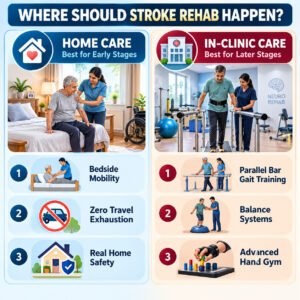
+-----------------------------------------------------------------------------------+
|                 COMPARING REHABILITATION DELIVERY MODELS                          |
+-----------------------------------------------------------------------------------+
| CLINICAL FEATURE       | HOME CARE PHYSIOTHERAPY      | IN-CLINIC NEURO-REHAB     |
+------------------------+------------------------------+---------------------------+
| Best Recovery Stage    | Months 1 & 2 (Severe fatigue,| Month 3 Onward (Upright   |
|                        | bed-bound, travel difficulty)| mobility, advanced gait)  |
+------------------------+------------------------------+---------------------------+
| Primary Focus          | Bed mobility, safe transfers,| High-tech gait retraining,|
|                        | real home safety adaptations.| parallel bars, hand-gym.  |
+------------------------+------------------------------+---------------------------+
| Patient Energy Cost    | Low (Zero travel fatigue).   | Moderate-High (Travel     |
|                        |                              | energy requirement).      |
+------------------------+------------------------------+---------------------------+
| Equipment Available    | Portable FES, resistance     | Full parallel bars, body- |
|                        | bands, balance pads, ramps.  | weight suspension systems.|
+-----------------------------------------------------------------------------------+
Advantages of Home Care Physiotherapy (Months 1 and 2)
During the first two months post-stroke, traveling in a car or auto-rickshaw through busy traffic causes severe physical fatigue, heightened sensory overload, and increased muscle spasticity.
Utilizing specialized home care physiotherapy in Bangalore offers critical advantages:
Real-World Environmental Assessment: The visiting physiotherapist inspects the patient’s actual living space—evaluating bed height, mattress firmness, door width, bathroom entry thresholds, and lighting.
Custom Home Modifications: Therapists assist families in identifying where to install stainless-steel grab bars, non-slip rubber mats, and ramps.
Family Caregiver Training: Caregivers are trained directly in their own home environment on how to perform safe transfers (bed-to-wheelchair, wheelchair-to-toilet) without injuring their own backs.
Advantages of In-Clinic Neuro-Rehabilitation (Month 3 Onward)
As the patient gains upright endurance and stability in Month 3, transitioning to our outpatient clinics in Banashankari, Basavanagudi, or Century Club unlocks advanced therapeutic resources:
Dedicated Rehabilitation Equipment: Access to gait-training parallel bars, body-weight supported treadmill systems, wall-mounted pulleys, and specialized hand-gym equipment.
Peer Support and Social Motivation: Training alongside other stroke survivors in a professional setting boosts psychological morale, reduces depression, and increases exercise compliance.
Detailed Clinical Case Study: 90-Day Transformation at Rayara Kirana
Patient Profile
Patient Name: Mr. Venkatraman R.
Age: 64 years old
Residence: Basavanagudi, Bangalore
Clinical Diagnosis: Right-sided hemiparesis resulting from an acute left middle cerebral artery (MCA) ischemic stroke.
Co-Morbidities: Type 2 Diabetes Mellitus, Essential Hypertension.
Baseline Status at Initial Evaluation (Day 10 Post-Stroke)
Neurological State: Severe right-sided flaccidity. Upper extremity score on Fugl-Meyer Assessment was 12/66; lower extremity was 18/34.
Mobility: Complete dependence. Bed-bound, requiring assistance from two adults for rolling and sitting transfers.
Postural Balance: Severe rightward leaning during attempted sitting; unable to maintain edge-of-bed posture for more than 30 seconds.
Psychological Status: High anxiety, emotional lability, and pronounced fear of remaining permanently bedridden.
The 12-Week Multi-Phase Recovery Plan
Phase 1: Months 1 (Weeks 1–4) — Foundation & Trunk Stabilization (Home Care)
Frequency: 6 days per week, 60-minute daily home visits by a senior neuro-physiotherapist.
Interventions: Passive and active-assisted range of motion for right upper and lower limbs. Daily FES applied to the right deltoid to prevent shoulder subluxation. Segmental rolling drills and gluteal bridging. Mirror-guided trunk centering exercises on the bed edge.
Month 1 Outcome: Achieved independent edge-of-bed sitting balance for 15+ minutes. Shoulder joint intact with zero subluxation. Successfully performed bed-to-chair transfers with single-person assistance.
Phase 2: Month 2 (Weeks 5–8) — Standing, Spasticity Control & Parallel Bars (Home Care + Clinic Transition)
Frequency: 5 days per week.
Interventions: Transitioned to standing weight-bearing. Introduced prolonged stretching and NDT handling to manage emerging spasticity in the right calf and flexor wrist. Fitted with a rigid thermoplastic Ankle-Foot Orthosis (AFO) to eliminate foot drop. Initiated parallel bar stepping drills.
Month 2 Outcome: Achieved 50/50 symmetrical standing weight distribution. Able to step forward and backward inside parallel bars with minimal manual guidance.
Phase 3: Month 3 (Weeks 9–12) — Functional Independence & Community Re-Integration (In-Clinic)
Frequency: 4 days per week at Rayara Kirana Basavanagudi Outpatient Center.
Interventions: Advanced gait training transitioning from parallel bars to a single-point quad-cane. CIMT protocol applied to the right arm (2 hours daily mitt wear on the left hand). Stair navigation retraining using the “Up with the good, down with the bad” methodology. Dynamic balance board training.
Month 3 Outcome: Independent ambulation using a quad-cane over indoor and outdoor flat surfaces. Berg Balance Scale score reached 48/56 (low fall risk). Re-established pincer grip to pick up objects and hold a coffee mug. Successfully navigated 15 steps to reach his second-floor residence.
Frequently Asked Questions About Stroke Physiotherapy
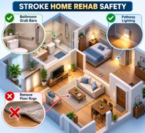
Practical Home Safety & Environmental Modification Checklist
To prevent secondary falls and support functional independence during the sub-acute phase, families should audit their home environment using the checklist below:
Bedroom Modifications
Ensure bed height allows the patient’s feet to rest flat on the floor when sitting on the edge.
Install a firm, supportive mattress that prevents sinking during edge-of-bed sitting.
Position a stable nightstand on the patient’s healthy side for easy access to water, glasses, and call bells.
Install motion-sensor nightlights along the pathway from the bed to the bathroom.
Bathroom Modifications
Install wall-mounted stainless-steel grab bars alongside the toilet and inside the bathing area.
Replace smooth floor tiles with non-slip textured rubber flooring or rubber safety mats.
Use a sturdy, height-adjustable shower chair to allow safe seated bathing.
Raise toilet seat height using an elevated toilet seat extension to simplify sit-to-stand transfers.
Hallways and Living Areas
Remove all loose throw rugs, carpets, and trailing electrical cords throughout the house.
Ensure clear, wide pathways between bedrooms, living rooms, and dining areas.
Secure firm armchairs with high backrests; avoid deep, soft sofas that make standing up difficult.
Install wooden handrails along both sides of indoor staircases.
Take Action: Schedule Your Comprehensive Neuro-Rehab Consultation
The first 3 months following a stroke represent a vital opportunity for physical and neurological recovery. Early, structured rehabilitation helps maximize gains during this high-neuroplasticity window.
Whether you require compassionate, specialist home care physiotherapy during the initial recovery phase or advanced in-clinic gait retraining, Rayara Kirana Physiotherapy & Rehab Center is committed to supporting your family through every step of the recovery journey.
Clinic Branches: Banashankari Stage II | Basavanagudi | Century Club, Bangalore
Home Visit Service Area: South Bangalore (Banashankari, Basavanagudi, JP Nagar, Jayanagar, Padmanabhanagar, Giri Nagar)
Lead Clinician: Dr. Kiran S. Murthy, PT (25+ Years Clinical Experience)
Book Your Appointment: Visit our Book Appointment page or contact our clinical care coordinator directly at +91-99727 77111 to schedule a comprehensive neuro-rehabilitation evaluation today.